Spectral domain optical coherence tomography for detecting glaucoma
Spectral domain optična koherentna tomografija za odkrivanje glavkoma
DOI:
https://doi.org/10.18690/actabiomed.98Keywords:
spectral domain optical coherence tomography, retinal nerve fiber, layer thickness, macular thickness, detecting glaucomaAbstract
Purpose: To evaluate the diagnostic ability of macular thickness parameters and peripapillary retinal nerve fiber layer (RNFL) thickness parameters for detecting glaucoma using spectral domain optical coherence tomography (SD–OCT).
Methods: 37 eyes of 20 glaucoma patients and 30 eyes of 16 healthy subjects included in this study underwent macular and peripapillary RNFL scans with SD–OCT using standard scanning parameters. The ”Macular Cube 512x128” scan protocol was used to measure the macular thickness. The ”Optic Disc Cube 200x200” scan protocol was used for assessing the peripapillary region. The discrimination power of all parameters for detecting glaucoma was determined by the Area under Receiver Operating Characteristics (AROC) curve and sensitivity at fixed specificity, followed by a comparison of the best macular thickness and peripapillary RNFL thickness parameters.
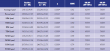
Results: Significant differences between glaucoma patients and healthy subjects were found in all peripapillary RNFL thickness parameters and all macular thickness parameters, except in the fovea (p=0.322). The largest AROC with sensitivity at >90% specificity among the peripapillary RNFL thickness parameters was found for the average peripapillary RNFL thickness (AROC 0.95, sensitivity 76%). The largest AROC with sensitivity at >90% specificity among the macular parameters was found for the inferior inner macular thickness (AROC 0.90, sensitivity 78%). There was no statistically significant difference between the AROCs of these two parameters (p=0.208).
Conclusion: To discriminate glaucoma patients from healthy subjects using SD–OCT, macular thickness parameters had high diagnostic ability which was comparable to that of the peripapillary RNFL thickness parameters.
Downloads
References
Downloads
Published
Issue
Section
License
Copyright (c) 2014 Acta Medico-Biotechnica

This work is licensed under a Creative Commons Attribution 4.0 International License.