Imaging, virtual reconstruction and computational material tissue) testing
Slikovna obdelava, virtualna rekonstrukcija in računalniško testiranje gradiv (tkiv)
DOI:
https://doi.org/10.18690/actabiomed.10Keywords:
imaging, virtual reconstruction, computational analysis, biological tissues, metallic foamsAbstract
Purpose: Recent advances in professional software and computer hardware allow for reliable computational analyses of new engineering materials as well as biological tissues. Therefore, the purpose of this paper is to describe the procedures allowing detailed reconstruction and virtual testing of such materials.
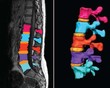
Methods: This paper describes the procedures and techniques for computational reconstruction of specimens, based on three-dimensional (3D) imaging data sets. First, different techniques of acquiring 3D imaging data sets (i.e., computed tomography – CT, magnetic resonance imaging – MRI and ultrasound – US) are introduced. Next the virtual reconstruction procedures for generated material (tissue) scans, based on image recognition, are addressed. For this purpose the up-to-date commercial software package ScanIP was used, allowing for an automatic virtual reconstruction.
Results: The reconstructed models can be virtually redesigned and adopted for special requirements or can be discretized (using +ScanFE and +ScanCAD) for further computational analysis, for example to predict their behaviour under quasi-static or dynamic loading conditions.
Conclusions: The paper concludes with three practical examples: (i) reconstruction and structural analysis of the proximal femur (alone and with a modelled implant), (ii) reconstruction of the lumbar spine and (iii) reconstruction and structural analysis of an irregular aluminium cellular material. The proposed procedures proved to be sophisticated and effective techniques suitable for a wide spectrum of medical and engineering applications.
Downloads
References
Downloads
Published
Issue
Section
License
Copyright (c) 2009 Acta Medico-Biotechnica

This work is licensed under a Creative Commons Attribution 4.0 International License.